

Daniel A. Lichtenstein
Ambroise-Pare Hospital, France
Title: The potential of lung ultrasound to diagnose hemodynamic pulmonary edema (the BLUE-protocol)
Biography
Biography: Daniel A. Lichtenstein
Abstract
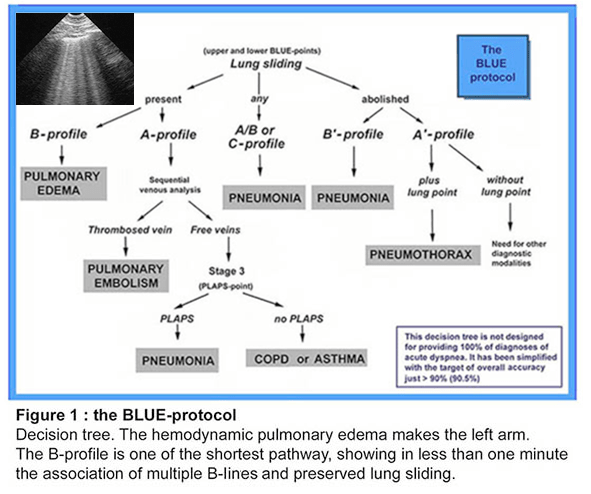
The diagnosis of hemodynamic pulmonary edema, sometimes difficult, usually requires irradiating techniques or indirect approaches (echocardiography, BNP), which are time-consuming. The BLUE-protocol proposes lung ultrasound as a direct diagnosis. What is required is a simple, gray-scale unit; a wide range (up to 17 cm) micro convex probe, the best for scanning together lungs and veins (and heart plus whole body). The B-line is the basis for the diagnosis, a certain kind of comet-tail artifact strictly defined for avoiding confusions. The B-profile is defined, in a supine or semi-recumbent patient, by anterior, bilateral and multiple B-lines, associated with a conserved lung sliding. This profile is called the B-profile. For the diagnosis of hemodynamic pulmonary edema, regardless any echocardiographic data, regardless the quality of a cardiac window, the B-profile has a sensitivity of 97%, and a specificity of 95%. The BLUE-protocol should be used first in a dyspneic patient, in order to see the reality of the pulmonary edema. Then, the patient can be treated accordingly. A test showing the origin of this edema is asked in a second step, calling an expert echo-cardiographist. The BLUE-protocol also allows, by an exclusive scanning of the lung and (when required) the veins, an accurate diagnosis of the other most frequent acute lung diseases: pneumonia, pulmonary embolism, exacerbated COPD, severe asthma and pneumothorax, using 7 other profiles (called A-profile, A’-profile, B’-profile, A/B-profile, C-profile). These profiles allow frequently or confidently differential diagnoses of hemodynamic pulmonary edema, such as pneumonia, pulmonary embolism, COPD. All in all, when using the BLUE-protocol wisely, i.e., surrounded by the clinical information and a few, basic tests, the diagnosis, positive or negative, of hemodynamic pulmonary edema is usually done confidently. Many subtleties (associated lung diseases, difficulties of diagnoses using too modern equipment (which destroy artifacts) are not dealt within this volume.
References:
- (2008) Lung ultrasound in acute respiratpry failure : the BLUE-protocol. Chest 134:117-125.
- (1998). A lung ultrasound sign allowing bedside distinction between pulmonary edema and COPD: the comet-tail artifact. Intensive Care Medicine 24:1331-1334
- (1994) Diagnostic échographique de l’œdème pulmonaire (Lettre à la rédaction). Revue d’Imagerie Médicale 6:561-562.
- (2016) Lung Ultrasound in the Critically Ill – the BLUE-protocol. Springer-Verlag, Heidelberg, pp 181-185
- (2004) van der Werf TS, Zijlstra JG. Ultrasound of the lung: just imagine. Intensive Care Med 30:183-184